Background
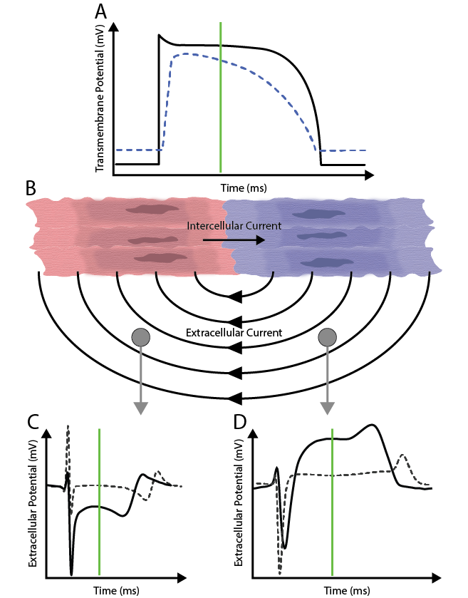
Myocardial ischemia is a disease of the heart characterized by an imbalance between nutrient supply and demand to the heart tissue. Myocardial ischemia increases the risk of arrhythmia and chronic myocardial infarction. Within ischemic areas of the heart, cellular action potentials are altered, leading to shorter, slower, and less stable electrical function. These electrical abnormalities can manifest on a body-surface electrocardiogram (ECG) as ST-segment elevations and depressions due to the formation of injury currents. Unfortunately, the ECG as a clinical marker for myocardial ischemia remains limited, with past studies showing poor sensitivity and specificity. In addition, body-surface recordings can only infer the locations of bioelectric sources, which can be crucial if surgical intervention is necessary.
Methods
At the CEG, we use advanced experimental techniques to characterize the three‑dimensional pattern of myocardial ischemia. We mapped ischemia‑related ST‑segment potential shifts during the acute phase of brief ischemic episodes produced by reducing coronary blood flow and increasing cardiac workload. These studies were conducted in open‑chest, intact pre-clinical models using intramural needle electrodes in combination with epicardial surface electrodes.

Our findings indicate that acute ischemia produces a complex electrocardiographic response, marked by a nonuniform pattern of ST‑segment elevation throughout the myocardial wall. Gaining a clearer understanding of the spatial and temporal behavior of the underlying diseased tissue could improve future approaches for localizing ischemia from cardiac surface measurements and may ultimately influence how body‑surface potentials are interpreted.