Electrocardiographic Imaging
Background
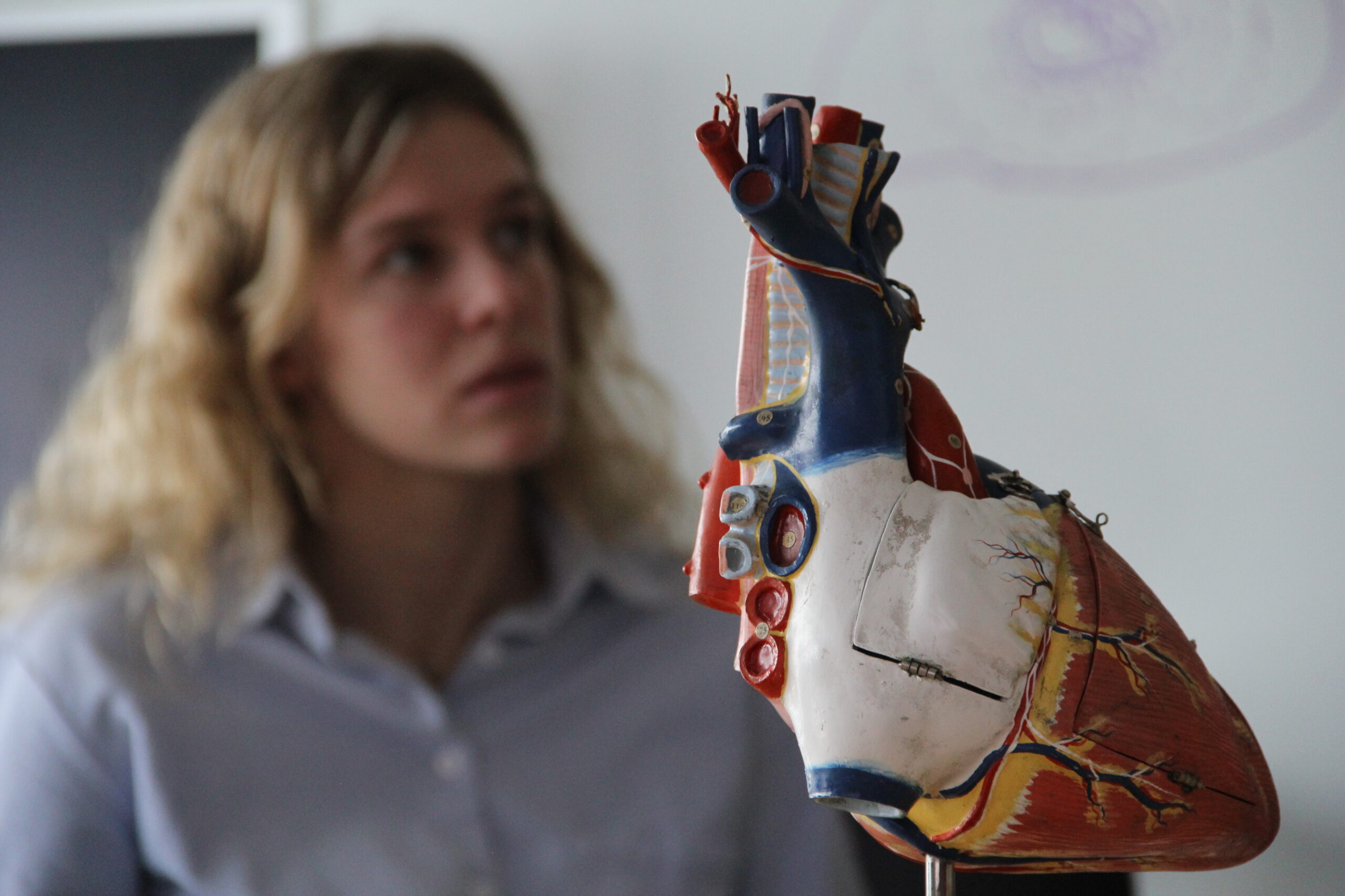
Electrocardiographic imaging (ECGI) is a technique that leverages the mathematical and biophysical relationships between the signals generated by the heart (the bioelectric source) and remote measurements such as those taken from the body surface. By understanding the forward relationship from heart bioelectric signals to torso surface measurements, we can solve for the inverse relationship: estimate bioelectric sources using measurements from the body surface. ECGI has a long history of development and application in scientific and medical domains. The CEG has a long history of innovations and contributions in this space including a strong presence in the consortium for electrocardiographic imaging (CEI, co-founded by Dr. Rob MacLeod).

Like most reconstructions, ECGI requires the solution to what is known as an inverse problem, a generalized formulation with applications across many diverse fields, including medical imaging, seismology, and astronomy. ECGI is the specific reconstruction of cardiac bioelectric activity from observed body surface potential signals. ECGI formulations require the solution of an inverse problem that is generally based on an associated forward problem, as depicted in the figure above. he forward problem describes the generation of body surface ECG signals from a known cardiac source model. Forward problems are common in many domains, with many modeling problems being themselves forward problems. For example, we can consider a scenario where we have a light bulb in the center of a room and a light sensor mounted on one wall of that room. In this case, we can define our forward problem as describing the brightness of light on a wall as measured by our sensor (a distant measurement point) resulting from the light bulb at the center of the room (the signal source.) To establish a forward relationship which would allow us to estimate how much light hits the wall, we would need to know various details about the system such as the distance between, and orientation of, the light and the wall. These relative locations constitute a geometric model for our light-wall system. We would also need a model of our light source, complete with relevant aspects such as the intensity of the light and what wavelengths the light bulb emits. Finally, we would have to incorporate some assumptions about the physics that dictate how light passes through the air and is reflected or absorbed by the wall, as well as any other physical attribute of our light-wall system. With a mathematical description of this physical model, and all of these inputs, we could then formulate a relationship between the light source and light measurement. From this forward relationship we could then compute the light intensity we would expect to measure at our wall-mounted light sensor. In a similar manner, the electrocardiographic forward problem also requires a source model, a geometric model, a physics-based formulation to tie the two together, and a mathematical formulation to compute a solution. In this case, we describe a bioelectric source and then incorporate the volume through which current passes (the volume conductors) to compute body surface potentials.
Methods
A wide variety of inverse solution methodologies exist to solve the ECGI inverse problem.

Relevant Papers
R. MacLeod and M. Buist, “The forward problem of electrocardiography,” in Comprehensive Electrocardiology (P. Macfarlane, A. van Oosterom, O. Pahlm, P. Kligfield, M. Janse, and J. Camm, eds.), pp. 247–298, London, UK: Springer Verlag, 2010.
A. Pullan, L.K.Cheng, M. Nash, D. Brooks, A. Ghodrati, and R. MacLeod, “The inverse problem of electrocardiography,” in Comprehensive Electrocardiology (P. Macfarlane, A. van Oosterom, O. Pahlm, P. Kligfield, M. Janse, and J. Camm, eds.), pp. 299–344, London, UK: Springer Verlag, 2010.